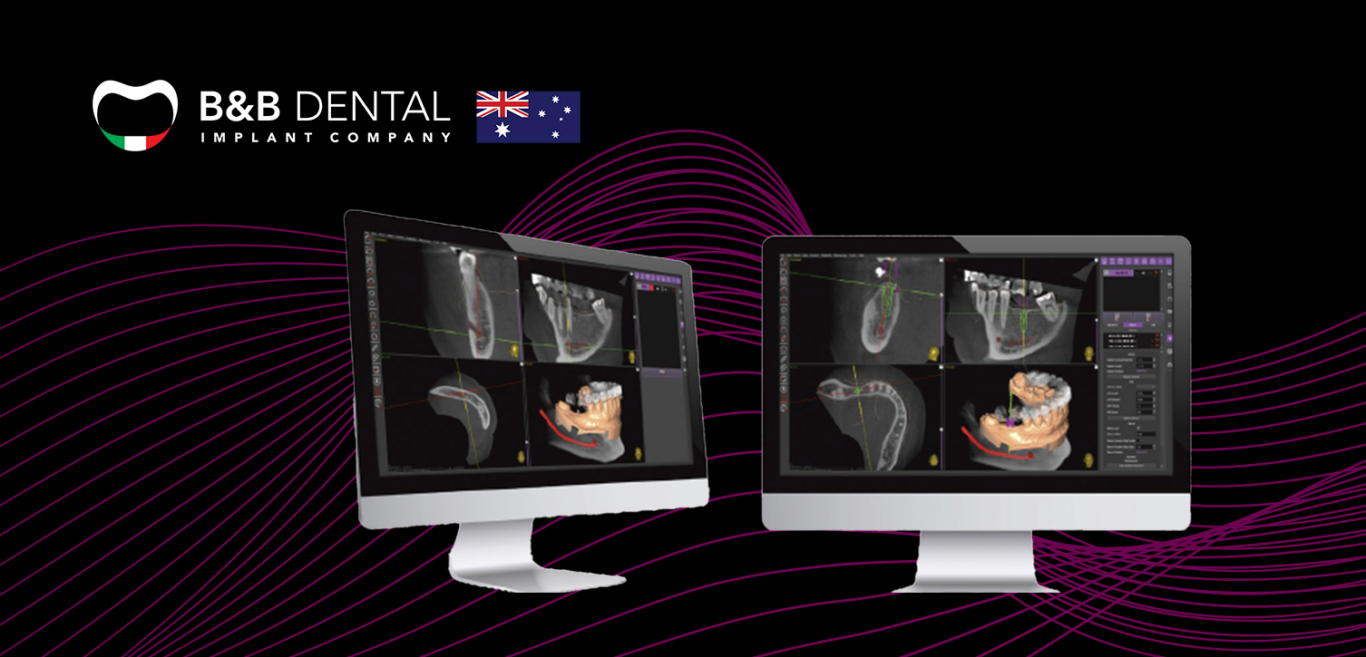
Surgical Guide Planning
1. DIAGNOSIS
To obtain a precise and usable image from X-rays, impressions, and intraoral scans, it's essential to configure the parameters accurately and ensure the proper positioning of the guide.
2. RADIOGRAPHIC TEMPLATE
For edentulous patients or those with significant metal restorations, a radiographic template aligns DICOM files and models. During Cone Beam, verifying mouth opening, bone thickness, tooth spacing, and checking for metal interference are crucial. Impression accuracy is key, with bicomponent silicones for partial edentulism and polyether for full edentulism.
3. DATA UPLODING
Patient data must be digitised for software comprehension:
- Extracting bone structure details from the obtained DICOM file.
- Capturing soft tissues and teeth using the STL of the scanned model.
- Establishing spatial alignment through the radiographic template, accurately positioned in the mouth during scanning and on the model itself.

